Research at Paul Strickland Scanner Centre is helping not just our own patients but those around the globe
Not long ago, being diagnosed with advanced head and neck cancer meant not only radiotherapy and chemotherapy, but usually also neck surgery – just to make sure the treatment had worked. Lymph nodes (a type of gland that forms part of the body’s immune system) were cut out and looked at under a microscope in a laboratory to make sure no cancer cells remain. The surgery can be painful, leave scarring, occasionally damage nerves resulting in “shoulder droop”, and there is a risk of death as in all major surgery. And even without complications, surgery is expensive to perform and puts a strain on the healthcare system.
Doctors had no choice but to advise surgery
In more than two-thirds of people, the neck surgery revealed that the treatment had been successful and the person was now cancer-free. However because it was the only sure way of telling whether someone could be given the all-clear, doctors had no choice but to advise surgery. Until recently, that is, when advances in scanning technology made a growing number of clinicians question whether there isn’t another way that is kinder, and evidence of an alternative way forward began to mount. “Before the advent of PET-CT it was impossible to tell which people had active disease and which ones didn’t,” says Dr Wai-Lup Wong, a consultant radiologist who works at Paul Strickland Scanner Centre. Dr Wong has been providing clinical leadership to our PET-CT team for many years. Amongst many other national key roles he chairs the group of doctors who advise NHS England on PET-CT.
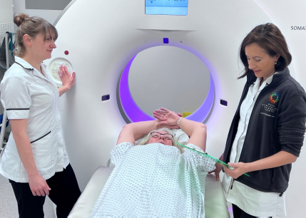
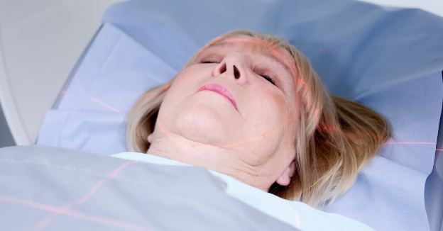
A PET-CT scan is a type of scan where people are injected with radioactive glucose, which is then rapidly taken up by cancer cells. Medical staff can then readily see where exactly in the body cancer is active, helping to make your treatment more successful. During 2016 we carried out more than 4,300 PET-CT scans at Paul Strickland Scanner Centre.
A large study was needed
Although some doctors suspected that a clear PET-CT scan might give a patient the “all-clear” as well as a biopsy could, there was just not enough evidence, and doctors just couldn’t be sufficiently confident to risk a patient’s life. What was necessary was a large study that provided evidence medical teams could rely on.
So it was in 2007, after receiving a grant from the National Institute for Health Research (NIHR), that Dr Wong and a number of clinicians launched the PET-Neck study, which aimed to see if PET-CT could reduce the number of unnecessary surgeries in patients with advanced disease. In what researchers call a “randomised controlled trial”, doctors asked people who had finished their treatment for advanced head and neck cancer whether they were interested in taking part in the study: a total of 564 at 43 hospitals throughout the UK (including all head and neck cancer treatment centres) volunteered, with half receiving neck surgery and the other half having a PET-CT scan. Where the PET-CT scan showed a patient had cancer, they went on to have the usual surgery, and doctors kept a careful eye on those patients where their scan had shown them to be in remission. Both groups were carefully monitored by their medical team during follow-up appointments for as long as 5 years to see how they did.

Clear results
The results were clear. “The study reduced unnecessary surgery by about 80% in patients who took part. Follow-ups showed that patients who had not had neck surgery but a PET-CT scan instead did just as well as those patients who had neck surgery – in other words, PET-CT spared this group of people head and neck surgery without having reduced their survival,” said Dr Wong.
Paul Strickland Scanner Centre played a vital role in the research, with Dr Wong taking on the role of PET-CT lead and co-principal investigator. His colleague Dr Bal Sanghera, our physicist, ensured quality control for the study. This was hugely important, as all the data had to be comparable across the UK and therefore all scans were centrally reviewed here. They then had to be sent back to every patient’s doctor in a timely fashion, so that those patients who had a scan that confirmed cancer could be treated swiftly.
International game-changer
Now practice is changing internationally and patients who have had chemotherapy or radiotherapy for advanced head and neck cancer will be offered a PET-CT instead, with doctors explaining to them that if the PET-CT scan is negative they are highly unlikely to have active disease in their neck. Dr Wong said: “Thousands of people throughout the NHS are now being spared unnecessary surgery. And an analysis found that it also saved the NHS money.”
Dr Wong said: “Thousands of people throughout the NHS are now being spared unnecessary surgery. And an analysis found that it also saved the NHS money.”
The results of the PET-CT Surveillance versus Neck Dissection in Advanced Head and Neck Cancer study have been published in the New England Journal of Medicine.